
| Scan type | EEG | CT | MRI | fMRI | PET |
|---|---|---|---|---|---|
| Example | 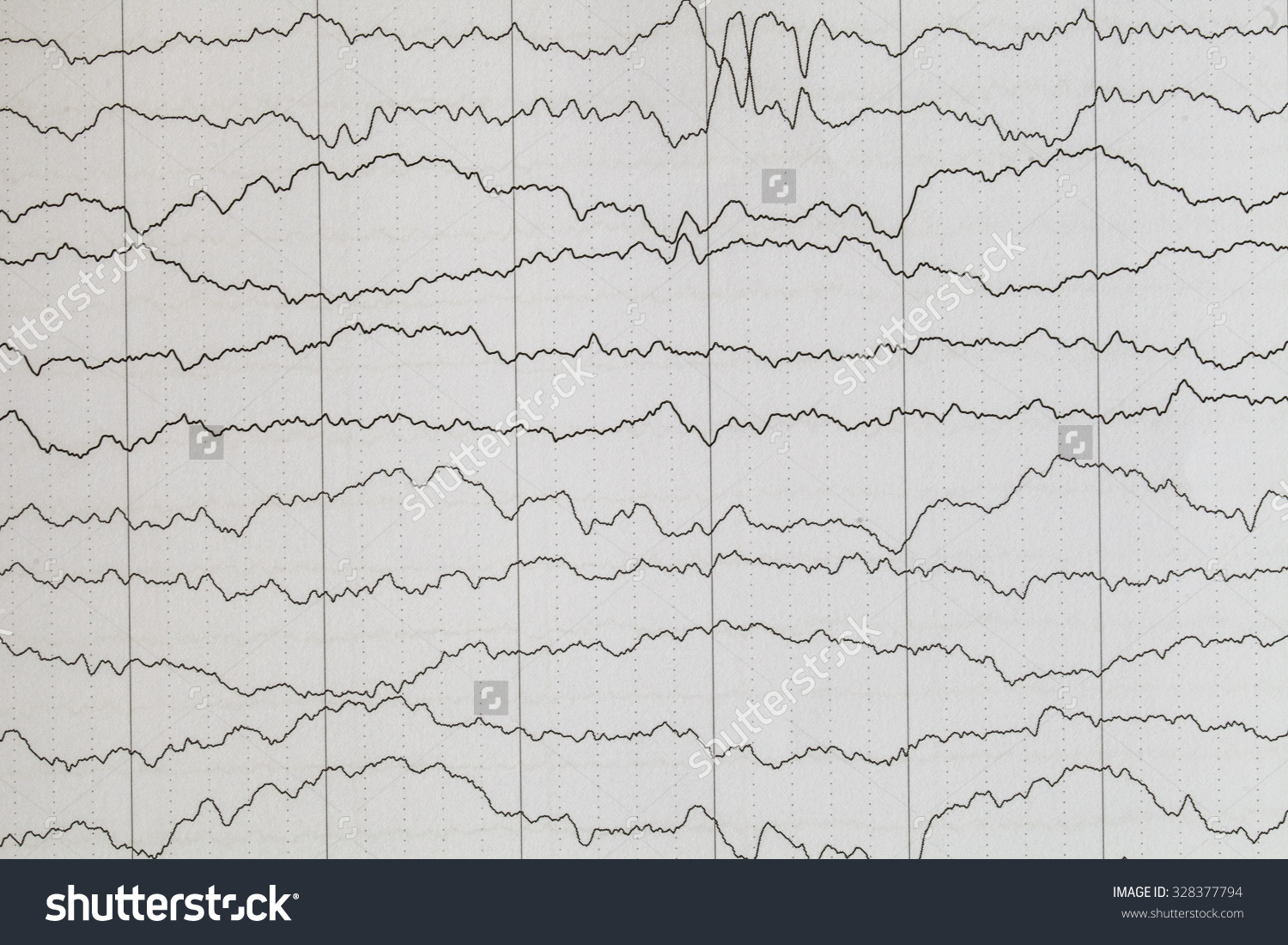 |
 |
 |
 |
 |
| Method | |||||
| Advantages | Detects biochemical changes in brain | ||||
| Disadvantages | Only shows activity in the cerebral cortex | High X-ray dose | Difficult for people with claustrophobia. | Difficult for people with claustrophobia. | |
| Some uses | Detection of brain injuries and skull fractures | Identify structures e.g. brain tumours, demyelinating nerve fibres, aneurysm | Imaging tumours |
Drag the Statements:
- Scalp electrodes detect voltage fluctuations
- X-rays show internal structure in slices, from any angle
- Strong magnetic field and a radio wave pulse. Protons in water give coloured 3D map of cortex
- A strong magnetic field and a radio wave pulse show flow of oxygenated blood
- FDG metabolism shows areas of glucose use
- Silent, non-invasive, does not use ionising radiation
- High resolution of bone, soft tissues and blood vessels at the same time
- Detailed anatomical image without using ionising radiation
- Assesses structure and functioning of e.g. the brain from second to second
- Exposure to γ-radiation
- Diagnosis of epilepsy
- Study brain function in real time