this involves the muscle producing tension and controlling the speed of the muscular contraction. This movement can be a concentric or eccentric muscular contraction.
Exercise Physiology
Chapter 2

This system is mainly concerned with producing movement through muscle contraction. This section explores the different types of muscles in our body and their involvement in sporting activities.
There are three types of muscle in the body:
Involuntary muscles are not under our conscious control which means we can’t make them contract when we think about it.
Voluntary muscles are under our conscious control so we can move these muscles when we want to.
There are three two different types of skeletal muscles:
Each type of muscle fibre has different characteristics which are shown in the table:
| Type I | Type II | |
|---|---|---|
| Speed of contraction | Slow | Fast |
| Force produced | Low | Medium/high |
| Resistance to fatigue | High | Medium/low |
| Colour | Red | White |
| Energy system | Aerobic | Anaerobic |
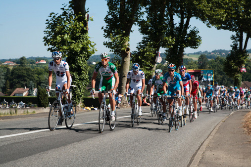 Cyclists need a high percentage of type I fibres so that their muscles can work for the duration of a race without getting tired. These muscles are red in colour because of the amount of capillaries that transport the oxygenated blood to the working muscles.
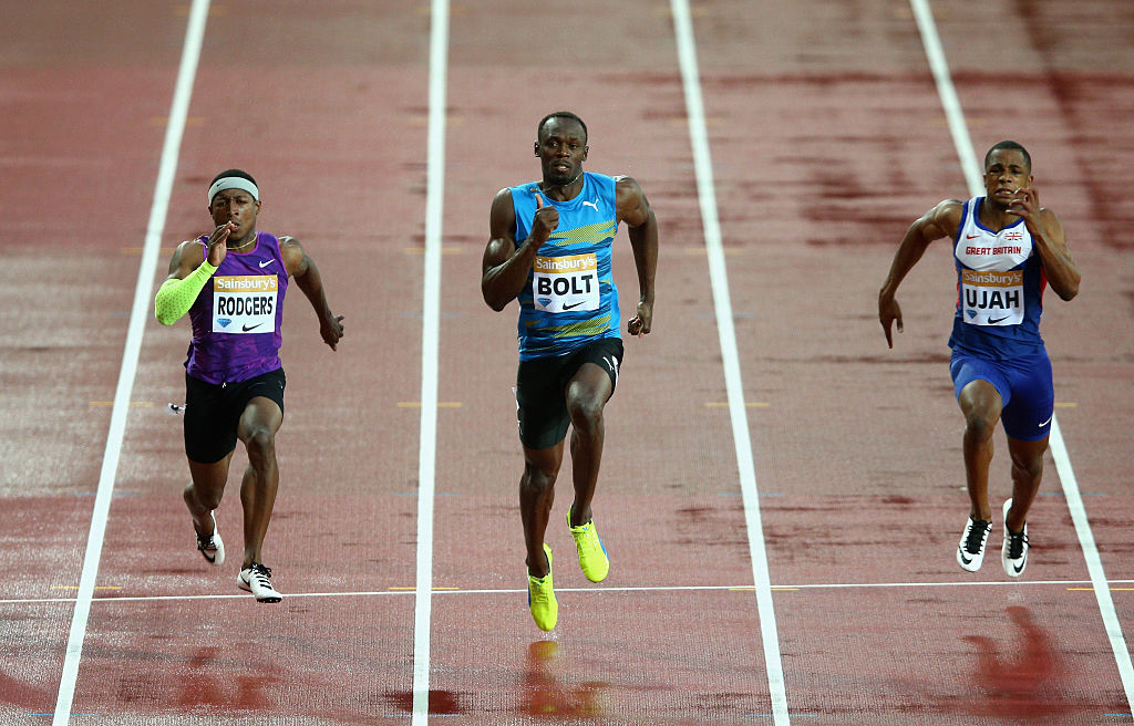 Sprinters need a high percentage of type II fibres which allow their muscles to contract very quickly. Fast muscle contractions give runners power and enable them to maintain a high speed over a 100 m distance. However, this type of muscle tires very quickly, meaning sprinters are not able to run at this speed for very long.
Explain which type of muscle fibre a long-distance runner requires in order to be successful in their event?
Muscles cause movement by contracting across joints. Muscles are attached to the skeleton by tendons in two places:
The origin is the end of a muscle which is attached to a fixed bone. The insertion is the other end of the muscle that is attached to the bone which moves.
Muscles contract in different ways to produce a range of movements:
this involves the muscle producing tension and controlling the speed of the muscular contraction. This movement can be a concentric or eccentric muscular contraction.
this involves the muscle shortening. The origin and insertion of the muscle move closer together and the muscle becomes fatter.
this involves the muscle lengthening whilst it is under tension. The origin and the insertion move further away from each other. An eccentric contraction provides the control of a movement on the downward phase and it works to resist the force of gravity.

Muscles are attached to bones by tendons. They move our bones and associated body parts by pulling on them – this process is called muscle contraction.
Because muscle contraction cannot push a bone back into its original position, muscles must work together in ‘antagonistic muscle pairs’. One muscle of the pair contracts to move the body part, the other muscle in the pair then contracts to return the body part back to the original position.
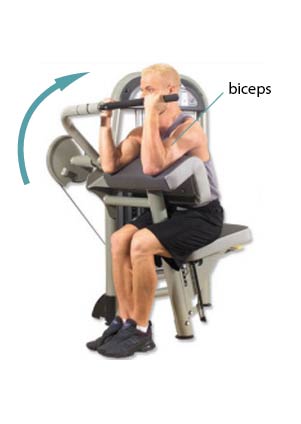
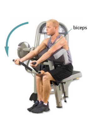
In an antagonistic muscle pair, as one muscle contracts, the other muscle relaxes. The muscle that is contracting is called the agonist and the muscle that is relaxing is called the antagonist.
Key fact: one way to remember which muscle is the agonist – it’s the one that’s in ‘agony’ when you are doing the movement as it is the one that is doing all the work!
For example, when you perform a bicep curl, the bicep will be the agonist as it contracts to produce the movement, while the tricep will be the antagonist as it relaxes to allow the movement to occur.
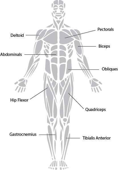
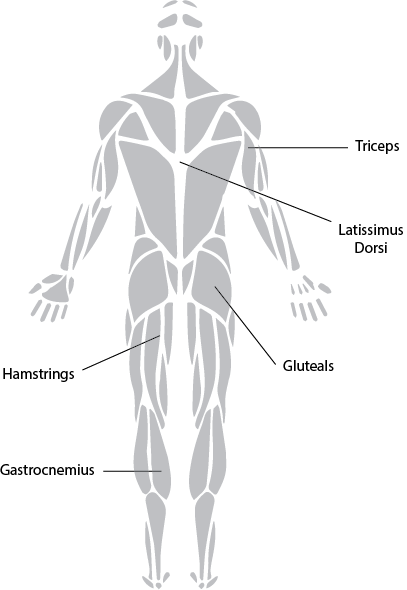
The following groups of muscles are antagonistic pairs:
| Biceps | Triceps |
| Hamstrings | Quadriceps |
| Gluteals | Hip Flexors |
| Gastrocnemius | Tibialis Anterior |
| Pectorals | Latissimus Dorsi |

Describe how the antagonistic muscle pairs are working at the elbow during the downwards and upwards phase of a press-up?
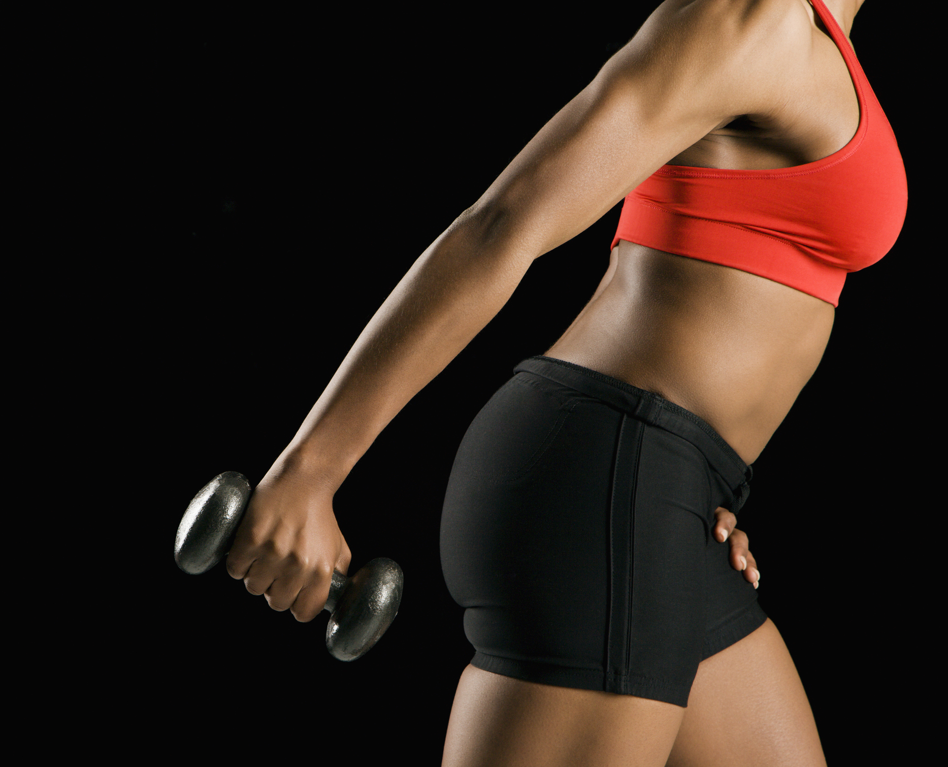 When a person takes part in resistance exercises such as weight training, their muscle tissue is placed under stress. This causes micro tears in the muscle fibres. The body responds by repairing the muscle fibres and making them larger.
When a muscle gets bigger, the process is called hypertrophy.
The skeleton is the central structure of the body and is made up of bones, joints and cartilage. The skeleton provides the framework for muscles and gives the body its defined human shape.
The main bones of the skeleton and their location are shown here:

The skeleton has five main functions:

These joints allow a wide range of movement and all have a similar joint structure.
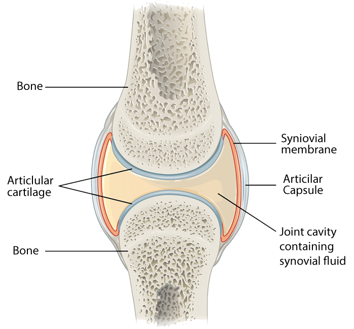
Different types of synovial joints allow varying degrees of movement, these include:

Which type of joint allows the greatest range of movement?
The different types of movement that are permitted at each joint are given specific terms.
Flexion – bending a joint. This occurs when the angle of a joint decreases. For example, the elbow flexes when performing a bicep curl.
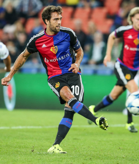
Extension – straightening a joint. This occurs when the angle of a joint increases, for example, when throwing a shot put.
Abduction – movement away from the midline of the body. This occurs in the hip and shoulder joints during a jumping jack movement.
Adduction – movement towards the midline of the body. This occurs in the hip and shoulder joints, returning the arms and legs back to their original position from a jumping jack movement.
Circumduction – this is where the limb moves in a circle. This occurs in the shoulder joint during an overarm tennis serve.
Rotation – this is where the limb moves in a circular movement around a fixed joint, towards or away from the midline of the body. This occurs in the hip joint in golf, while performing a drive shot.
| TYPE OF JOINT | BODY LOCATION | TYPES OF MOVEMENT |
|---|---|---|
| Ball and socket | Hip, shoulder | Flexion/extension, rotation, abduction, adduction, circumduction |
| Hinge | Knee, elbow | Flexion/extension |
| Pivot | Neck | Rotation |
The muscular system works in conjunction with the skeleton to produce movement of the limbs and body.
Ligaments and tendons are two main types of connective tissue that help the muscular-skeletal system produce movements.
The muscles contract to pull on the bones to produce movements. Joints are able to move in a variety of directions to allow us to perform a range of sporting movements.
An analysis of a netball shot shows how the muscular-skeletal system works together to produce this throw.

| Joint | Type of movement | Bones | Muscles | Muscle contraction | |
|---|---|---|---|---|---|
| Phase 1 | Elbow | Flexion | Humerus, Radius, Ulna | Biceps, Triceps | Concentric |
| Phase 2 | Elbow | Extension | Humerus, Radius, Ulna | Triceps, Biceps | Concentric |
Complete the table to analyse a rugby conversion kick.

| Joint | Type of movement | Bones | Muscles | Muscle contraction | |
|---|---|---|---|---|---|
| Phase 1 | Knee | ||||
| Phase 2 | Knee |
The cardiovascular system is made up of three main parts and consists of the heart, the blood vessels and the blood that flows through them.
If you clench your hand into a fist, this is approximately the same size as your heart. It is located in the middle of the chest, slightly towards the left.
The heart is a large muscular pump and is divided into two halves - the right-hand side and the left-hand side.
The right-hand side of the heart is responsible for pumping deoxygenated blood to the lungs.
The left-hand side pumps oxygenated blood around the body.
Each side of the heart consists of an atrium and a ventricle which are two connected chambers.

The atria (plural of atrium) are where the blood collects when it enters the heart.
The ventricles pump the blood out of the heart, to the lungs or around the body.
The septum separates the right and left-hand side of the heart.
The tricuspid valve is located between the right atrium and right ventricle to prevent backflow of blood from the ventricle to the atrium.
The bicuspid valve is located between the left atrium and left ventricle to prevent backflow of blood from the ventricle to the atrium.

There are four main blood vessels that take blood into and out of the heart.
The aorta takes blood from the left ventricle to the body.
The vena cava takes blood from the body back to the heart.
The pulmonary artery takes blood from the right ventricle to the lungs.
The pulmonary vein returns blood from the lungs to the left ventricle.
How to remember the facts – it’s a letter game:
arteries = away (from the heart)
The main artery is the aorta
The main vein is the vena cava
The cardiovascular system has three main functions:
When blood is pumped out of the heart, the blood vessels open (vasodilation) to allow the large volume of blood to leave the heart. In order for the blood to reach the working muscles, some of the other blood vessels close (vasoconstriction). This process also helps to return the blood back to the heart (venous return) and to remove the waste products.
In the heat, blood vessels close to the surface of the skin enlarge. This process is called vasodilation. This allows more heat to be lost from the blood.
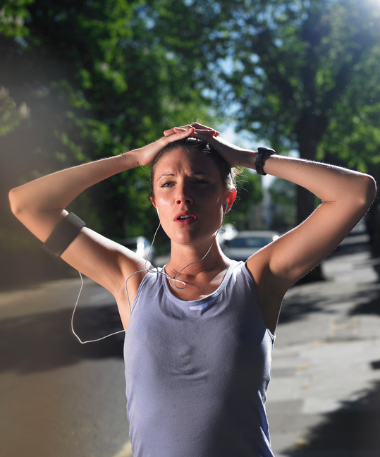
When a person takes part in exercise, their face can become pink due to vasodilation of the blood vessels close to the skin’s surface.
In the cold, blood vessels at the skin’s surface close. This process is called vasoconstriction and takes blood away from the surface of the skin to help prevent it from losing heat.
Cardiac output (Q) is the amount of blood pumped from the heart every minute and is the product of heart rate (HR) and stroke volume (SV).
Heart rate (HR) is the number of times your heart beats in one minute. The average number of beats is 72 beats/minute.
Stroke volume (SV) is the volume of blood pumped out of the heart each beat. The average amount of blood/beat is 0.7 litres.
| Cardiac Output = Stroke volume x Heart rate |
|---|
| Q = SV x HR |
| 4.9 litres/minute = 0.07 litres x 70 beats/minute |
The fitter you are, the larger your stroke volume and the lower your heart rate, therefore your cardiac output stays the same.
During exercise, tidal volume increases as the depth and rate of breathing both become greater. This has the effect of taking more oxygen into the body and removing more carbon dioxide.
| Measure | Rest | Moderate Exercise |
|---|---|---|
| Heart rate | 72 beats/minute | 120 beats/minute |
| Stroke volume | 0.07 litres | 0.2 litres |
| Cardiac output | 5.04 litres/minute | 24 litres/minute |
When the heart contracts, it pushes blood into the blood vessels, creating blood pressure.
A blood pressure reading consists of two values:
Systolic is when the heart contracts and diastolic is when the heart relaxes.
The average blood pressure for an adult is 120/80 mmHg. The first number is the systolic value and the second number is the diastolic value.
Blood pressure is determined by Q (cardiac output) and the resistance to the blood flow (R). Resistance to blood flow is caused both by the diameter of the blood vessels and by the thickness of the blood.
Any changes to heart rate, stroke volume and cardiac output are determined by the intensity and duration of exercise.
Heart rate is measured in beats per minute (bpm). During exercise the heart rate increases so that sufficient blood is taken to the working muscles to provide them with enough nutrients and oxygen. An increase in heart rate also allows for waste products to be removed.
Maximal heart rate can be worked out by the following equation:
| Maximum HR = 220 – age |
|---|
What is the maximum HR of a 16-year-old person?
Stroke volume increases which means more blood is pumped out of the heart every time it contracts.
At rest, a person’s cardiac output is approximately 5 litres per minute. During exercise, this can increase to as much as 30 litres per minute as both the heart rate and stroke volume increase.
Work out the cardiac output of a person at rest with a HR of 70 bpm and an SV of 70 ml. Compare that to their cardiac output when they are taking part in exercise as their HR increases to 120 bpm.
When taking part in exercise, there is a greater cardiac output because the athlete requires more blood and oxygen to be transported to the working muscles. The increase in the amount of blood also helps with the removal of waste products such as lactic acid and carbon dioxide.
A typical blood pressure reading for a person at the start of exercise would be around 160/85 mmHg.
The main role of the respiratory system is to transport oxygen from the air that we breathe through a system of tubes into our lungs and then into the blood stream.

Gaseous exchange occurs in the alveoli in the lungs and takes place by diffusion.
Diffusion is the movement of gas from an area of high concentration to an area of low concentration.
In the alveoli, there is a high concentration of oxygen and in the bloodstream, there is a high concentration of carbon dioxide.
Oxygen diffuses into the blood from the alveoli and carbon dioxide diffuses into the alveoli from the blood.

At the muscle, the opposite occurs and carbon dioxide enters the blood from the muscle and oxygen enters the muscles.
Capillaries surround the alveoli in the lungs. Both the capillaries and alveoli walls are very thin – just one cell thick. They are made of semi-permeable membranes which allow oxygen and carbon dioxide to pass through them.
Describe the process of gaseous exchange at the muscles.
Vital capacity is the maximum amount of air that can be breathed out after breathing in as much air as possible. Taking part in regular aerobic exercise has been shown to increase a person’s vital capacity.
Breathing rate (frequency) is the number of breathes in a minute. The average breathing rate is 12 breaths/minute.
Tidal volume is the amount of air breathed in with each normal breath. The average tidal volume is 0.5 litres (500 ml).
Minute ventilation (VE) is the total volume of air entering the lungs in a minute. The average minute ventilation is 6 litres/minute.
| Minute ventilation = Breathing rate x Tidal volume |
|---|
| VE = BR x TV |
| 6 litres/minute = 12 x 0.5 |
During exercise, tidal volume increases as the depth of breathing increases and the rate of breathing increases too. This has the effect of taking more oxygen into the body and removing more carbon dioxide.
| Measure | Rest | Moderate Exercise |
|---|---|---|
| Breathing rate | 12 breaths/minute | 30 breaths/minute |
| Tidal volume | 0.5 litres | 3 litres |
| Minute ventilation | 6 litres/minute | 90 litres/minute |
The cardiorespiratory system works together to get oxygen to the working muscles and remove carbon dioxide.
During exercise, the muscles need more oxygen in order to contract and they produce more carbon dioxide as a waste product. To meet this increased demand by the muscles, the following happens:
Breathing depth (tidal volume) and rate increase – this gets more oxygen into the lungs and removes more carbon dioxide out of the lungs.

The graph shows that as a person goes from rest to exercise, their tidal volume increases.
Heart rate increases – this increases the rate that oxygen is transported from the blood to the working muscles and carbon dioxide is transported from the working muscles to the lungs.
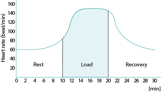
This graph shows that:
Depending upon whether the body uses oxygen or not in order to perform specific exercises determines if the exercise is aerobic (with oxygen) or anaerobic (without oxygen).
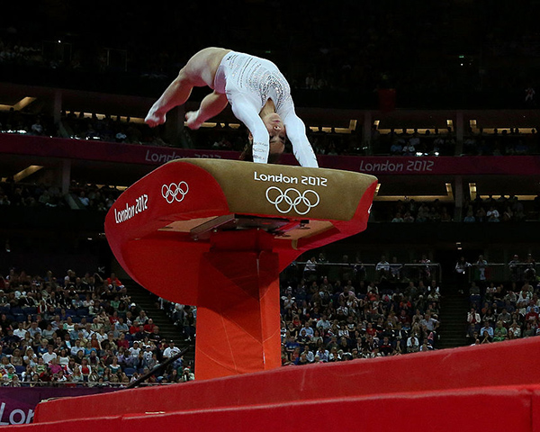
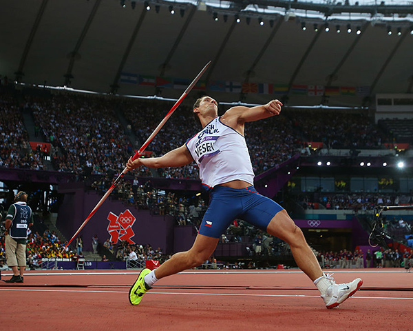
The anaerobic respiratory system supplies energy very quickly for sports such as vaulting in gymnastics or throwing a javelin where the activity only lasts a few seconds.
There are two types of anaerobic energy systems. These are:
The creatine phosphate (CP) anaerobic system supplies energy faster than all other energy systems.
It is used for explosive, high-intensity contractions, such as in sprinting 100 metres, but it can only supply energy for about ten seconds.
| CP → energy + creatine |
|---|
Once the CP system has run out, the lactic acid system is used to supply energy.
This system breaks down glucose into lactic acid. It produces energy very quickly, but not as quickly as the CP system.
| Glucose → energy + lactic acid |
|---|
The lactic acid energy system produces the majority of the energy for moderate to high intensity activities such as running 400 metres.
Which sport would use the creatine phosphate energy system?
Which sport would use the lactic acid energy system?
It is the lack of oxygen and the build up of lactic acid that causes fatigue.
Both these systems require oxygen to restore them which is called an oxygen debt.
After taking part in exercise, a person continues to breathe more deeply and rapidly than when at rest to take in additional oxygen to repay this oxygen debt.
The oxygen is then used to:
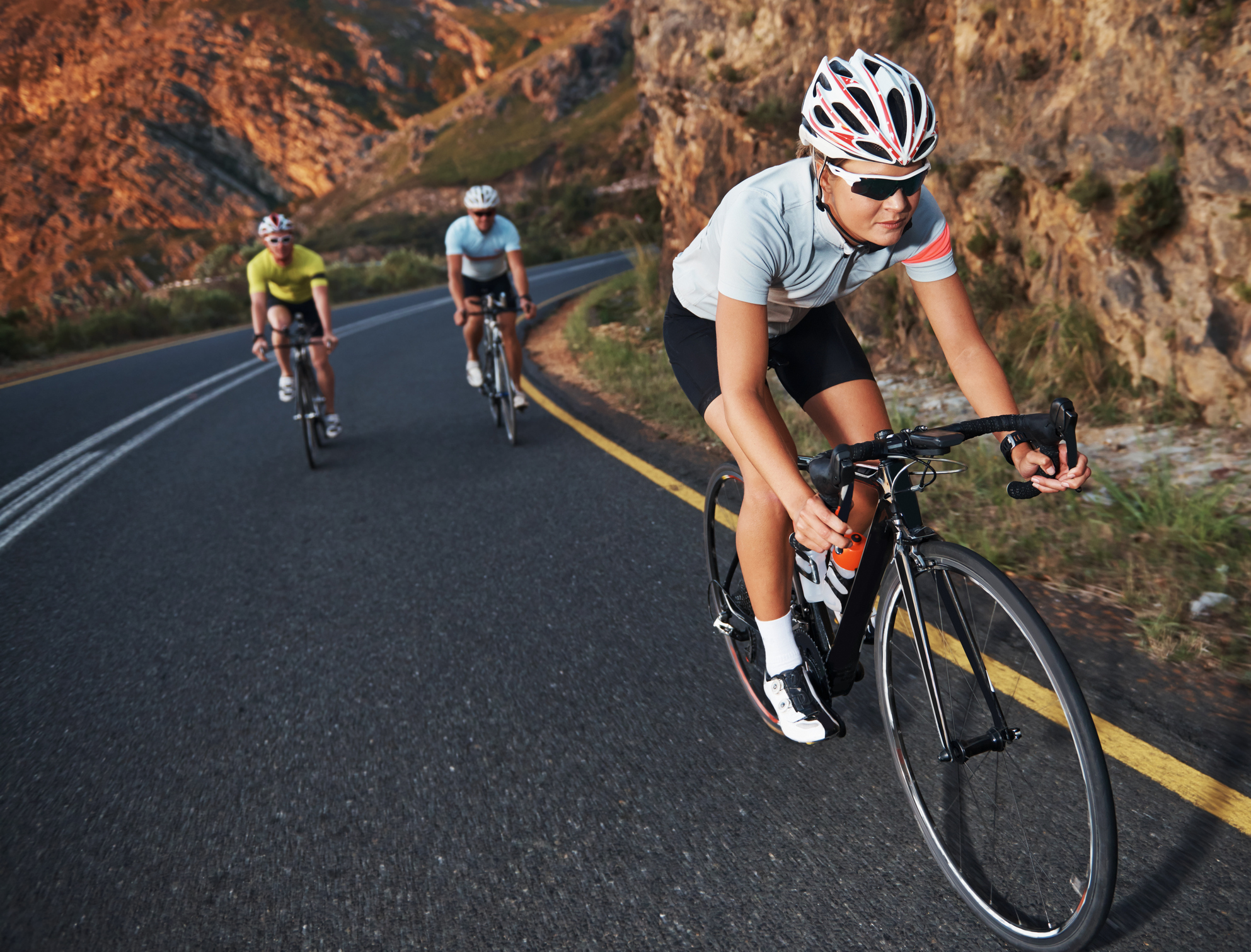
The aerobic respiratory system is responsible for producing the majority of our energy while our bodies are at rest or taking part in low-intensity exercise for long periods of time, such as jogging or long-distance cycling.
| Glucose + O2 → energy + H2O + CO2 |
|---|
Carbohydrates and fats supply the energy for the aerobic energy system and can supply energy for long periods of time.
Carbohydrate food sources include rice; bread; potatoes; bananas and energy drinks. Fat food sources include butter; oils; cheese; milk and nuts.
Give an example of an athletics event that mainly uses the aerobic energy system to provide energy.
The aerobic respiratory system is responsible for producing the majority of our energy while our bodies are at rest or taking part in low-intensity exercise for long periods of time, such as jogging or long-distance cycling.
When a person takes part in exercise, their body systems provide energy for these activities. After regular exercise participation, these systems adapt to improve exercise performance.
When a person takes part in exercise the cardiovascular, respiratory, energy and muscular systems all work together to supply energy to the working muscles and remove waste products.
| Short-term effects of exercise | |
|---|---|
| Cardiovascular system | Increase in stroke volume (SV) Increase in heart rate (HR) Increase in cardiac output (Q) Increase in blood pressure (BP) |
| Respiratory system | Increase in breathing rate Increase in tidal volume |
| Cardio-respiratory system | Increase in oxygen uptake Increase in carbon dioxide removal |
| Energy system | Increase in lactate production |
| Muscular system | Increase in temperature of muscles Increased pliability Muscle fatigue |
After exercising, the muscles need to rest, adapt and recover. There is a risk of injury if the body is not rested for long enough after exercise.
Why is there less chance of a person straining a muscle if they have completed a warm up session before taking part in exercise?
Taking part in regular exercise, around 3 times per week for 6 weeks, will lead to adaptation of the body systems that are trained. This has the effect of increasing performance in that type of exercise or sport.

| Long-term effects of exercise | Type of training | |
|---|---|---|
| Cardiovascular system | Cardiac hypertrophy Increased stroke volume (SV) Decrease in resting heart rate (HR) Increase in maximum cardiac output (Q) Capillarisation of lungs and muscles Increase in number of red blood cells |
Aerobic |
| Respiratory system | Increased vital capacity Increased number of functioning alveoli Increased strength of the respiratory muscles (internal and external intercostals and diaphragm) |
Aerobic |
| Energy system | Increased production of energy from the aerobic energy system Increased tolerance to lactic acid |
Aerobic Anaerobic |
| Muscular system | Muscle hypertrophy Increased strength of tendons Increased strength of ligaments |
Resistance |
| Skeletal system | Increase in bone density | Resistance |
The muscle wall of the left ventricle increases in size so is able to pump out more blood during each beat which increases the stroke volume. As the stroke volume is increased, the resting heart rate decreases but cardiac output (Q) remains the same as SV × HR = Q.
Capillarisation is the process where new capillaries are formed. Capillarisation takes place at the alveoli in the lungs and in skeletal muscle. This has the effect of increasing the amount of oxygen that can be transferred to the working muscles as well as increasing the amount of carbon dioxide that can be removed.
Give an example of a type of exercise that would produce cardiac hypertrophy.
Give an example of a type of exercise that would produce muscle hypertrophy.