Content
- Joints and articulations
- Planes and axes of rotation
- Movement patterns
- Levers
To understand how to improve technically the athlete or coach must understand the component parts of the technical model and the movements that contribute to successful performance
Knowledge of the skeletal system and how it works helps us to understand movement and explains how skills are performed. The possible movements at each joint or articulation (where to or move bones meet) can help coaches understand to skill development and performance improvement.
Skeletal joints are classified according to their structural and functional (range and type of movement) characteristics.
Joints (structural classifications):
Most common joint in the human skeletal system, they have a joint cavity and ligaments hold the articulating bones together. These joints are freely moveable.

Synovial Joint Structure
Types of Synovial Joints
| Synovial Joint | Structure | Movement | Example |
|---|---|---|---|
| Ball and socket | Tri-axial, most mobile | Flexion/extension, abduction/adduction, rotation, circumduction | Hip Shoulder |
| Hinge | Uni-axial, one plane | Flexion/extension | Elbow knee |
| Pivot | Uni-axial, one plane | Rotation | Neck - atlanto-axial |
| Gliding | Bones glide past each other | Carpals, tarsals | |
| Condyloid/ellipsoid | Bi-axial – two planes | Flexion/extension, abduction/adduction | Wrist – radio-carpsals |
In order to understand sporting performance we need to be able to describe the movement of the human body. Using the planes and axes of movement and joint actions we can describe performance and understand the way people move.
Planes of Motion: There are 3 imaginary anatomical planes that intersect at the body’s centre of gravity dividing the body into equal portions

Most movements in sport occur in multiple planes. However, some movements are more planar (in one plane) than others.
When movement is in one plane, it means that no part of the body crosses from one side of the plane to the other during the movement.
There are 3 imaginary anatomical axes that intersect at the body’s centre of gravity. In most sporting actions, movement is about more than one axis

Using the planes and axes of movement and joint actions we can describe performance and understand the way people move.
| Joint | Movement | Plane | Axis | Example |
|---|---|---|---|---|
| Hinge – Elbow | Flexion/extension | Sagittal | Transverse | Biceps curl |
| Ball and socket – Shoulder | Flexion/extension Flexion – arms up | Sagittal | Transverse | Line out jumping |
| Ball and socket - Shoulder | Abduction –away, Adduction-toward | Frontal | Frontal | cartwheel |
| Ball and socket – Shoulder | Circumduction – combination of movements | Frontal/Transverse | Frontal/Transverse | Bowling |
| Ball and socket - Shoulder | Rotation - twisting | Transverse | Longitudinal | Golf drive |
| Ball and socket - Shoulder | Horizontal abduction/adduction | Transverse | Longitudinal | Discus |
| Ellipsoid – Ankle | Dorsiflexion-toes up, Planterflexion - pointed | Sagittal | Transverse | Long jump or gymnastics |
| Ellipsoid – Wrist | Pronation – palm down, Supination – palm up | Sagittal | Frontal | Discus, Rolling ball |
Levers allow us to create movement that is greater than the force applied. The skeleton forms a system of levers that allow us to move.
Functions of levers:
Levers are made up from three components:
There are three classifications of lever:
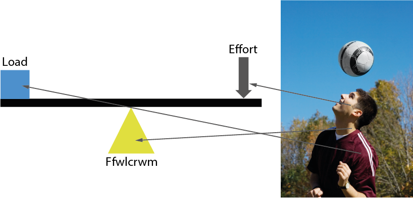
1st Order Lever e.g neck – heading the ball
2nd Order Lever e.g. ankle – planter flexion

3rd Order Lever e.g. knee kicking a ball
This is the most common lever in the human body and increases the body’s ability to move quickly, however it is inefficient at applying force.

Most levers in the body are 3rd class levers, this means: